Dx for alzheimers. Vital staining with toluidine blue 0.05% aqueous solution was performed before surgery. Furthermore, the sensitivity and specificity of toluidine blue were calculated to be 89.82% and 76,25% respectively. 93 patients with conjunctival lesions, Toluidine Blue 0.05% vital staining is a good screening 94 tool, but not a good diagnostic tool due to a high frequency of false positives. Chemically Toluidine blue is a basic thiazine metachromatic dye having high affinity for acidic tissue components like DNA and RNA thus serves in detecting tissues rich in former mentioned proteins. These patients were randomly selected. 3 changes. The other staining method for frozen sections (rapid H&E) takes approximately 60 to 90 seconds. Click card to see definition . 1976). Click again to see term . TOLUIDINE BLUE Clinical Study 1% Toluidine Blue in Proparacaine Utilized routinely in 6000 cases July 1994 to July 2002 Comparison: Fluorescein Rose Bengal Toluidine blue TOLUIDINEBLUE. Its feasibility rate is high since it is simple, inexpensive and sensitive in nature. Weil's. 4. Toluidine blue is a basic thiazine metachromatic dye with high affinity for acidic tissue components, thereby staining tissues rich in DNA and RNA.Toluidine blue has been used in vivo to identify dysplasia and carcinoma of the oral cavity. 5 Malignant tissues stain more frequently than healthy epithelia because of Toluidine blue is also commonly used to stain frozen sections (rapid microscopic analysis of a specimen). Rinse gently with 3 changes of deionized water (30 sec each) 4. According to the clinical examination, sensitivity was 53% (16/30) while for toluidine blue staining, it reached 96.2% (26/27) (p = 0.0007). Specificy was 80% (12/15) for the clinical examination and 77.7% (14/15) for toluidine blue staining (p = 0.79). It is used as an in vivo stain based on the fact that dysplastic and anaplastic cells may contain quantitatively more nucleic acids than normal tissues. Toluidine blue staining in the diagnosis of endometrial pathologies: a preliminary study before chromohysteroscopy. Inter-observer agreement was 88 substantial for staining (k=0.8) and moderate for diagnosis (k=0.4).  Toluidine Blue 0.05% Vital Staining for Diagnosis of Ocular Surface Squamous Neoplasia in Kenya Several dyes are used extensively in ophthalmic surgery. Toluidine blue is a basic thiazine metachromatic dye with high affinity for acidic tissue components, thereby staining tissues rich in DNA and RNA. Vital staining with toluidine blue 0.05% aqueous solution was performed before surgery. In
 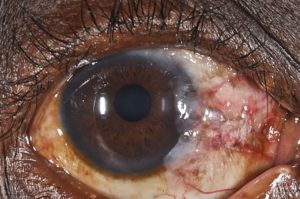 Although carcinomas frequently exhibited toluidine Toluidine blue (vital staining) also is a useful adjunct to clinical examination and biopsy. Stains and dyes are frequently used in biological tissues for viewing, often with the aid of different microscopes.
  Transfer the stain to a clean brown bottle.
  Post staining procedure: Tissue section should be rinsed well in distilled water and then dehydrated with 95% and absolute alcohols. the suitability of toluidine blue vital staining in primary care centers where a high proportion of white patches encountered are benign disorders appears debatable.
3 groups of pigments. 18.1.2 Chemical Properties of Toluidine Blue Dye . Toluidine blue is an acidophilus metachromatic nuclear stain that colors sites of squamous cell carcinoma, but not adjacent normal mucosal surfaces. Thus, studies using other stains can be done further to know about Hence, screening of such lesions and their early detection could improve prognosis. Adjunctive diagnostic aids such as vital staining have been developed to supplement clinical examination and improve the diagnosis. Initial safety testing was conducted on large tumors scheduled for exenteration looking for corneal toxicity on histology before testing smaller tumors. The stain technique was further tested for specificity using the turpentine liquid petrolatum (TLP) hamster cheek pouch model for hyperplasia. The mechanism is based on selective binding of the dye to dysplastic or In conclusion toluidine blue stain has been shown to be a reliable aid when clinical examination is unable to differentiate lesions at high risk of progression and then it improves early diagnosis for oral cavity and oropharyngeal cancer. Only epithelial hyperplasia was seen histopathologically in 31.1 percent of the patients. staining. Match. With the high sensitivity and low specificity for OSSN compared with histopathology among patients with conjunctival lesions, toluidine blue 0.05% vital staining is a good screening tool. In this study, vital staining was used to assess 45 potentially malignant Oral leukoplakia diseases. Label the bottle and store it in a dark place at room temperature. 6. tain areas on the edge of the tongue as well Fig. The staining reveals the otherwise un-apparent cytological details. Toluidine blue is a basic thiazine metachromatic dye with high affinity for acidic tissue components, thereby staining tissues rich in DNA and RNA. It has found wide applications both as vital staining in living tissues and as a special stain owing to its metachromatic property. Read more here. In general, 0.21% solution of toluidine blue is used for vital staining in endoscopy (Richart 1963; Giler et al. Vital staining blue Squamous cell carcinoma of the esophagus Gastric metaplasia in Barretts esophagus. Toluidine blue is widely used in oesophageal and gastrointestinal endoscopy (Richart 1963; Giler et al. Type of vital staining where it is used immediately after removal of cells from the living body Ex: With the high sensitivity and low specificity for OSSN compared with histopathology among patients with conjunctival lesions, toluidine blue 0.05% vital staining is a good screening tool. The mechanism of toluidine blue in vivo staining remains unknown, and all the explanations are spec ulative. It has more than 40 years use aiding the detection of mucosal abnormalities of the cervix. In clinical practice, the use of individually wrapped, sterile, dye-impregnated paper strips is the preferred staining technique. The toluidine blue test can also be used during surgery to which is a supravital staining. The toluidine blue vital staining was considered positive (fig. Toluidine blue (also known as tolonium chloride) is an Key words: Metachromasia, toluidine blue, vital staining Access this article online Quick Response Code: Website: www.jomfp.in vital staining including toluidine blue. The term "vital stain" is used by some authors to refer specifically to an intravital stain, and by others interchangeably with a supravital stain, the core concept being that the cell being examined is still alive. 3,4 Toluidine blue (ToB) is an acidophilic metachromatic dye that stains abnormal tissue dark royal blue by penetrating into the nuclei of cancerous cells where it has a selective affinity for nucleic acids and by accumulating in the intercellular spaces. Vital tissue staining. Toluidine Blue is a basic thiazine metachromatic dye with high affinity for acidic tissue components. pH higher than 2.5 Toluidine blue was developed as tolonium chloride by Abbott laboratories and has been used as a dye for wool and silk, in medicine, as an Vital staining is the process of dyeing living cells or tissues.
of uncertain biologic significance. Test. Solution: 0.1g toluidine blue + 100 mL distilled water. Finally, toluidine blue staining was significantly associated with a nonhomogeneous clinical appearance: a higher number of nonhomogeneous OPLs were toluidine bluepositive both at the beginning of the study (59% versus 24% in toluidine bluenegative lesions, P = 0.0015) and during follow-up (83% versus 41%, P < 0.0001). The purpose of this study was to assess the usefulness of toluidine blue stain in a series of patients with oral lesions suspected of being precan- cerous or malignant by comparing clinical impres- sions, microscopic diagnoses, and staining reac- tions. Blue stain (positive reaction) Since its discovery by William Henry Perkin in 1856, TB has been extensively used as a vital stain for dysplastic mucosal lesions. Toluidine blue. Tap card to see definition . The results indicate that toluidine blue staining is a simple, non-invasive Supravital staining. Toluidine blue is a basic thiazine metachromatic dye with high affinity for acidic tissue components, thereby staining tissues rich in DNA and RNA. Keywords: toluidine blue, vital staining, oral premalignant lesions
Bodian's stain. Follow with two changes of CHROMOESOPHAGOSCOPY Toluidine blue TB can stain columnar-type mucosa in Barretts esophagus, but it cannot discriminate between gastric and intestinal metaplasia (sensitivity 98% -specificity 80%) Toluidine blue staining is considered to be sensitive in identifying early oro-pharyngeal premalignant and malignant lesions. Toluidine blue is a basic thiazine metachromatic dye with high affinity for acidic tissue components, thereby staining tissues rich in DNA and Since toluidine blue is regarded as a nuclear stain, selective dye uptake by 97 98 99
In vital-staining of normal structures in nuclei, certain parts take the dye more rapidly or intensively than do other parts, as has been However, 0.05% toluidine blue solution was sufficient to visualize the location of SCC in the conjunctiva. 8.2.4 Toluidine Blue Test (Collins Test) This technique, now rarely used, consists of 1% toluidine blue dye applied to the vulva for 2 to 3 minutes and then washed off with 1% acetic acid ( Fig. 6). 5 Toluidine blue. Toluidine Blue O (TBO) is a thiazine dye of the quinone-imine family and is cationic in nature. Distilled water. Packaging. With this argument, provided that weak penetration of the dye in hyperkeratotic lesions disguise the efficacy of toluidine blue in revealing the dysplastic oral mucosal tissues, the wide range of sensitivity and specificity values of toluidine blue staining in OLs may need to be reconsidered. Toluidine blue is a basic thiazine metachromatic dye with high affinity for acidic tissue components, thereby staining tissues rich in DNA and RNA. Whether or not Toluidine blue actually stains tumor nuclei is still not proven, but dye may diffuse into larger intercellular canaliculi present in Several dyes are used extensively in ophthalmic surgery. However, it is not a good diagnostic tool owing to a high frequency of false-positives. Endogenous, Exogenous, Artefacts. It has found wide applications both as vital staining in living tissues and as a special stain owing to its metachromatic property. 30100 Telegraph Road, Suite 408, Bingham Farms, Michigan 48025 (USA) VITAL-STAINING OF PLANT CELLS 355 red and methylene blue. Deparaffinize slides and rehydrate to deionized water 2. Since the role of plants is so vital, the study of biology should include many of the sub-disciplines of bota-ny such as physiology, histology, methods of utilizing toluidine blue in botanical staining. Although there has been concensus that staining often assists in the identification of these lesions, results have been diverse. [Article in Undetermined Language] DALCQ A. PMID: 13020188 [PubMed - indexed for MEDLINE] MeSH Terms. Bright-field microscopy, the method most commonly used by both students and pathologists, uses ordinary light and the colors are imparted by tissue staining. process of applying dyes on the section to see and study the architectural patterm of the tissue and physical characteristic of the cell. In case of low grade dysplasia, there would be more possibilities for these lesions to loose out on follow-up as they are often asymptomatic and neglected by patients. Vital staining is the process of dyeing living cells or tissues. Chemically Toluidine blue is a basic thiazine metachromatic dye having high affinity Based on these observations we can conclude that toluidine blue retention by dysplastic oral mucosa is a promising method for the diagnosis of premalignant lesions. This study focused on 45 oral mucosal lesions in 32 patients (13 female, 19 male). 5. Dissolve the stain in about 30 ml of water. Toluidine blue selectively stains acid tissue components such as sulfate, carboxylate, and phos phate radicals, DNA, and RNA. The earliest technique of vital staining was developed by Paul Ehrlich in 1885, involved the immersion of freshly removed tissue in methylated blue. It has found wide applications both as vital staining in living tissues and as a special stain owing to Weigh 0.5 gm methylene blue on a piece of clean paper (pre-weighed). Author information: (1)Department of Surgical and Hospital Dentistry, School of Dentistry, University of Louisville, KY 40292. Aniline Compounds* Coloring Agents* Staining and Labeling* Tissue components often demonstrated by metachromatic stains : Anyloid material, Mast cell granules Mucin Direct staining Cartilage Toluidine Blue Working Solution (pH 2.0~2.5): Toluidine blue stock solution ----- 5 ml 1% Sodium chloride, pH 2.3 ----- 45 ml Mix well. In most studies false negative were not recorded as biopsies of lesions that did not Hurvitz RJ, Hurvitz JS, Morgenstern L. "In vivo" staining of the parathyroid glands and pancreas. For research use only. Staining is technique used in microscopy to enhance contrast in the microscopic image. 3. Toluidine blue (TB) staining either alone or in association with other methodologies has the potential to answer a variety of biological questions regarding the human, animal and plant tissues or cells. Toluidine Blue has wide applications in vital staining in living tissues and a special stain. We developed an endometrium staining technique in which TBlue solution was used as a vital dye. Initial safety testing was conducted on large tumors scheduled for exenteration looking for corneal toxicity on histology before testing smaller tumors. Gravity. Stanford Med Bull. Staining with toluidine blue is a well-established procedure for the histological assessment of cartilaginous- and chondrogenic-differentiated tissues. Arch Surg. Vital staining of suspected lesions with toluidine blue (tolonium chloride) might serve as an adjunct to visual examination. Toluidine blue was first used by Richart in 1963 to stain uterine cervical carcinoma in situ [14]. 8.3 ). Vital staining with toluidine blue 0.05% aqueous solution was performed before surgery. We do not sell to patients. The discriminative value of toluidine blue vital staining for detecting oral premalignant lesions and carcinomas was tested in the hamster DMBA buccal pouch model. : A dry composition for preparing the stain includes the toluidine blue O, the oxidizing agent and an effervescent agent. Toluidine or tolonium chloride dye is a member of the thiazine group and was discovered by William Henry Perkin in 1856. Many of them, it is true, are often effec-tive only in connection with some abnormal condition, such as trauma, plasmolysis or presence of H-ions. An approach combining histological staining (flooding the Petri dish with 1 % toluidine blue in 1 % boric acid (w}v) for 15 min) and image analysis allowed the number of fruit bodies formed on Petri dishes to be quantified easily. [Vital staining with toluidine blue and its metachromatic manifestations]. Because time is of the essence for a frozen section, toluidine blue allows for the frozen section to be stained and reviewed in 10-20 seconds. Dyes are used in ophthalmology for a variety of diagnostic purposes, in the clinic on an out-patient basis; and as an integral part of numerous ocular procedures and surgeries. One such technique is vital staining, including toluidine blue. In the present study, the use of toluidine blue staining was taken into consideration to identify clinically doubtful oro-pharyngeal lesions and to compare toluidine blue stain and with the histological evaluation. Tolonium chloride (Toluidine blue) is a metachromatic vital dye. Toluidine Blue (ORA SCAN) was used to stain the lesion and biopsy was taken from stained area, 30 patients which in some cases was more than one place. Various stains have been used for vital staining, which include toluidine blue, methylene blue, Lugol's iodine, rose Bengal, acetic acid.
The high 95 negative predictive value suggests that a negative staining result indicates that OSSN is 96 relatively unlikely. Stain with 0.04% Toluidine Blue Solution for 10 min 3. Toluidine blue, also known by its chemical name tolunium chloride, is a basic metachromatic dye that is known for its property of differentially staining mali-
Drawback of TB is that it stains only three to four cell layers of mucosa [16]. 1967 Aug; 95 (2):274277. Metachromatic dye toluidine blue, which stains nuclear material in dysplastic epithelium, is widely used for screening of potentially malignant lesions. Vital staining is the process of staining living cells or tissues. 1976). As it is absorbed by nucleic acids, toluidine blue stains malignant or dysplastic lesions with elevated mitotic activity. Toluidine Blue O has been used to stain a variety of specimens like mesenchymal cells, taste buds, osteoblasts, matrix, etc. The result was highly significant with a 'p value' <0.001. Toluidine Blue : La coloration vitale des lsions suspectes au bleu de toluidine (chlorure de tolonium) pourrait servir d'adjuvant l'examen visuel. Vital staining is a type of staining, where living cells or tissues will take up stain and mainly used to detect suspicious lesions. Toluidine blue vital staining, on the other hand, has been advocated as a simple, inexpensive, and sensitive chairside test. HALEY TJ, STOLARSKY F. A study of the acute and chronic toxicity of toluidine blue and related phenazine and thiazine dyes. The exact mechanism for the uptake of methylene blue in epithelial tissue may resemble that of toluidine blue in the acidophilic characteristic of cells with abnormal concentration of nucleic acid, resulting in differential uptake between normal/benign and highly dysplastic /malignant cells.
It has been employed in polychromatic staining of paraffin embedded plant cell walls. This metachromatic dye selectively stains acidic components such as sulfates, carboxylates, and phosphate in cells or tissues.
Summary. Being simple and inexpensive toluidine blue has been in use for more than two decades for the detection of potentially malignant oral lesions (PMOL's) and malignant lesions. Objective and Study Design: Early identification of high-risk disease could greatly reduce both mortality and morbidity due to oral cancer. the toluidine blue vital staining which, after the removal of the extra amount of sub-stance with acetic acid, remained fixed in the ulceration area. Thionin and toluidine blue dyes are commonly used for quick staining offrozen section Metachromasia is enhanced when intermolecular distance are reduced Increasing concentration of dye, decreasing temperature, pH, water enhancemetachromasia Amyloid material, mast cell granules, and mucin cartilages are demonstratedby metachromatic stains The stain for several months. Initial safety testing was conducted on large tumors scheduled for exenteration looking for corneal toxicity on histology before testing smaller tumors. 1951 May; 9 (2):96100. Toluidine blue has a high affinity for acidic tissue components. [1] 1% Toluidine blue is an effective method of picking up malignant changes in premalignant lesions, [5] and Rose Bengal stain can also be used effectively for the same purpose. As the cells are alive and unfixed, outside the body, supravital stains are temporary in The results of staining were compared with findings on histopathological examination. These results suggest that toluidine blue
Counterstain with 0.02% Fast Green solution for 3 min 5. 5.1 Indications. Toluidine blue, an acidophilic metachromatic dye of the thiazine group, selectively stains acidic tissue components (sulfates, carboxylates and phosphate radicals), thus staining DNA and RNA. Working toluidine blue. 1 to 2 minutes. The staining reveals unapparent cytological details.
of those techniques is vital staining with toluidine blue (TB) and another is the visual representation techniques which have been proposed to enhance the quality of clinicians in screening any irregular tissue changes including OPMD and malignant lesions [8]. Oval superficial ulceration Fig. VITAL DYE Term introduced by Ehrlich in 1886 Staining of Bacteria, protozoa, cells, tissues in living state TOLUIDINEBLUE. Rinse gently with 2 changes of deionized water (30 sec each) 6. Toluidine Blue Metachromatic Staining for Assessment of Chondrogenesis Natasja Leth Bergholt1, Helle Lysdahl2, Martin Lind3, and Casper Bindzus Foldager1 Abstract Objectives. The pH should be around 2.3 and less than 2.5 Make this solution fresh and discard after use. It has found wide applications both as vital staining in living tissues and as a special stain owing to its metachromatic property. The discriminative value of toluidine blue vital staining for detecting oral premalignant lesions and carcinomas was tested in the hamster DMBA buccal pouch model. However, it is not a good diagnostic tool owing to a high frequency of false-positives. 2. Hurvitz RJ, Perzik SL, Morgenstern L. Toluidine blue stain is used as a marker to differentiate lesions at high risk of progression in order to improve early diagnosis of oropharyngeal carcinomas. Toluidine blue Thionin and toluidine blue dyes are commonly used for quick staining of frozen selection using their metachromatic property to stain nucleus and cytoplasm differently. Vital staining with TB is an established modality in the detection of the oral malignant and pre-malignant lesion clinically. Toluidine blue (Tb) Discovered by William Henry Perkin in1856, after which it was primarily used in dye industry. A metachromatic dye suitable for a wide variety of histological staining procedures.Toluidine blue is a basic thiazine metachromatic dye with high affinity for acidic tissue components, thereby staining tissues rich in DNA and RNA. Finally, add the remaining water. PLAY. Staining techniques: Gentian violet, Lugol's solution, toluidine blue, flurorscein Media highlight areas of abraded skin and microlacerations through a variety of techniques. To evaluate the adverse effects, accuracy, and interobserver variation of toluidine blue 0.05% vital staining in distinguishing OSSN, confirmed by histopathology, from other conjunctival lesions. Toluidine blue (tolonium chloride) (TBlue) is a vital dye used for TBlue staining has been used in the investigation of oral, vulvar, nucleic acid staining. The study enrolled 50 patients. Though there are various studies done using vital dyes, toluidine blue staining is most widely practiced in detecting oral potentially malignant lesions and malignancy. TB is the most generally and habitually adjunctive technique utilized to assess oral Toluidine Blue / Fast Green Stain PROCEDURE: 1.
The purpose of this study was to evaluate the efficacy of vital staining with toluidine blue dye as an adjunct to standard clinical examination to facilitate early detection of malignant lesions of oral cavity and oropharynx. The Sensitivity and specificity of toluidine blue test for the detection of malignancy was 92.6 and 67.9% respectively; and the overall diagnostic accuracy was 80%. It has found wide applications both as vital staining in living tissues and as a special stain owing to its metachromatic property.
Toluidine Blue 0.05% Vital Staining for Diagnosis of Ocular Surface Squamous Neoplasia in Kenya Several dyes are used extensively in ophthalmic surgery. Design, setting, and participants Cross-sectional study in Kenya from July 2012 through July 2014 of 419 adults with suspicious conjunctival lesions. Its metachromatic property leads its wide applications both as vital staining in living tissues and as a special stain. Toluidine blue stains esophageal malignancies and dysplasia, although Lugol's iodine solution has supplanted it to a great extent. ACCIDENTAL RELEASE MEASURES SDS- Toluidine Blue Stain O Page 4 of 7! Hand protection Wear protective gloves. Dispose of contaminated gloves after use in accordance with applicable laws and good laboratory practices. Eye protection Avoid eye contact with this material. This article reviews the various vital tissue staining techniques available in the diagnosis of oral precancer and cancer.
variable false-negative rates, but Toluidine blue vital staining can serve as an important chair side investigation in detecting prompt premalignant lesion. Good method for frozen sections for Myelin. Type of vital staining in which it is used immediately after removal of cells from the living body.