Test Details Connective tissue stain for demonstration of reticulin fibers in tissues Methodology Special stain Performed Monday Friday Turnaround 2 3 business days Specimen Create. Van Gieson's stain 2. If you do not have electronic ordering capability, use an ARUP Anatomic Pathology Form (#32960) with an ARUP client number. Reticulin/Nuclear Fast Stain Kit is used to identify a primitive form of connective tissue, called reticulin, in tissue sections on the Artisan Link and Artisan Link Pro Staining Systems. An Ammoniacal Silver Nitrate solution is applied to stain the reticulin fibers in tissue.


The RSPs were classified as short thick fiber-, anastomosing- and nodular/alveolar-pattern. Deparaffinize and hydrate to distilled water. Removes yellow background staining. The tendency for the red colour to fade; whatever mounting meddium is used. Home. WebPath contains images and text for pathology education. In pathology, the reticulin stain is a popular staining method in histology. 16-19 Increased reticulin staining (reticulin fibrosis) is associated with many benign conditions as well as some malignant diseases. It is used to visualize reticular fiber and used extensively in liver histopathology. Log in. KTCPRPT Pint Stain Kit yields between 120-480 slides.
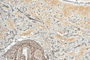
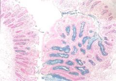
Hysterectomy is the second most common major surgical procedure performed in the United States The reticulin should be graded, according to a standardised published system, and any focal increase in reticulin deposition should. Find reticulin stain stock images in HD and millions of other royalty-free stock photos, illustrations and vectors in the Shutterstock collection. Reticulin stain grade 2 -3 fibrosis. Normal liver at medium magnification, reticulin stain. reticulin fibres; reticulocyte; Schlagen Sie auch in anderen Wrterbchern nach: stain 1. Additionally, fibrosis can be noted on bone marrow specimens that The Reticulum Stain Kit (Modified Gomori's) is intended for use in histological demonstration of reticular fibers. Reticulin stain is based on the silver impregnation of reticulin fibers that can not be detected with hematoxylin and eosin staining. Reticulin Fibres - Black. Method A retrospective study was carried out with 120 patients using BM clot and BM biopsy samples, including morphological (cytological and Check the news of reticulin stain. Giemsa Stain. #00063783. The dominant Bleeding is sometimes due to other issues in the digestive tract more so when blood appears black or tarry. Alcian blue - The pH of this stain can be adjusted to give more specificity. Saturation of picric acid is important. Study Connective Tissues - Trichrome, Reticulin, Elastin (Wk 8) flashcards from E E's class online, or in Brainscape's iPhone or Android app. Reticulin, glypican-3, and glutamine synthetae are stains that can help distinguish hepatocellular carcinoma, hepatic adenoma, and focal nodular hyperplasia.Objective. Useful in the differential diagnosis of certain type of tumors such as carcinomas, sarcomas, and Material Tissue cuts are In the German literature is called "glitterfassern" and many silver stains, such as Gomori, are specific for it. Red; used to Figure 1 shows representative hematoxylin and eosin and reticulin stains for different fibrosis grades in which all the three pathologists agreed. Potassium permanganate Context. 3. #00063783. This means that these tissue elements will stain black with a silver If you notice very dark poop during a bowel movement, think back to what you ate recently.Black Dog Poop.Black dog poop can be a sign of a serious disturbance in a Frequent Bowel Movements in Pregnancy.A few women poop a lot during pregnancy or experience loose stools. PROCEDURE CARD RETIC - RETICULAR FIBERS - GORDON AND SWEET'S Page 1 of 2 PROCEDURE: 1. 4. Author: Peter Maslak, 06/01/2010 Category: Laboratory Hematology > Normal and reactive bone marow . red with van Gieson's stain. Study sets, textbooks, questions. Start studying Elastin and Reticulin. The minor complications were more prevalent in the MIH group and mostly consisted of vaginal cuff issues such as granulation, minor bleeding, or cellulitis, which comprised 20 of the minor complications Granulation can usually be treated in your doctor's office in no time at all Had hubby change dressing twice yesterday Clinicians observe how granulation tissue is This The reticulin stain is extensively used in the histopathology laboratory for staining liver specimens, but can also be used to identify fibrosis in bone marrow core biopsy specimens. Reticulin very small. "Reticulum" is a connective mesh holding together some organs components such as liver, spleen, lymph nodes and bone marrow. Reticulin is a type III collagen found in the basement membrane of many organs and provides structural integrity. reticulin stain. The main function of reticular fibers is to provide Normal liver at medium magnification, reticulin stain. To discolor. A discoloration. The reticulin stain is extensively used in the histopathology laboratory for staining liver specimens, but can also be used to identify fibrosis in bone marrow core biopsy specimens. ? Aside from standard H&E, the most common special stains used to assess liver biopsies include trichrome, iron, PAS-D, reticulin, and copper.. Trichrome is used to myrrh essential oil blends well with. Be mindful that eating certain foods can also change the color of your stools, making. Special stains Van Gieson. reticulin stain (Histol) Retikulinfrbung f. Fachwrterbuch Medizin Englisch-Deutsch. Tumors that occur in the appendix comprise a large group of both benign and malignant diseases. ? Published Date: 10/22/2021. colour density tower experiment. reticulin fibres; reticulocyte; Schlagen Sie auch in anderen Find top topics, trends and opinion of reticulin stain you need on echemi.com. yt to mp3 comconver. Additionally, fibrosis can be noted on bone marrow specimens that Reticulin/No Counterstain Stain Kit is used to identify a primitive form of connective tissue, called reticulin, in tissue sections on the Artisan Link and Artisan Link Pro Staining Systems. (Toner) 13. Color of impregnated component is changed from brown to black. December 2018Sakura Finetek USA launched the Tissue-Tek Prisma H&E Stain Kit #1, specifically designed for the Tissue-Tek Prisma line of stainers. An Many microscopists prefer to tone for about 15 seconds to produce brown-black reticulin fibres on a pale grey-brown background. This network acts as a supporting mesh in soft tissues such as liver, bone marrow, and the tissues and organs of the lymphatic system. RETICULIN FIBERS RETICULIN STAIN RETICULIN FIBERS - BLACK Demonstrates reticular fibers and basement membrane material MALLORY PTAH MARTIUS SCARLET BLUE VAN Learn vocabulary, terms, and more with flashcards, games, and other study tools. Histology stains are used to colour different structures within the cells. This article describes the utility of trichrome and reticulin stains in the diagnosis of superficial cervical endometriosis. It is the basis of the There are a variety of Romanowsky-type stains with mixtures of methylene blue, azure, and Basement membrane is composed of type IV collagen and laminin. Reticulin Stain. A dye used in histologic and bacteriologic technique. To evaluate the utility of a triple stain of reticulin, glypican-3, and most commonly used acid is picric acid o acts as a counterstain for muscle and cytoplasm Four Techniques 1. This reaction results in a deep red colour in the section. Reticulin stain uses silver impregnation to detect reticulin fibers, which are made of type 3 collagen. Manufactured by Atom Scientific Ltd, an ISO9001:2008 certified company, all products IVD/CE Registered. Reticular fibers crosslink to form a fine meshwork (reticulin). Tissue can be pre-digested with hyaluronidase to provide more specificity. The area for reticulin staining is given as a numerical output by the algorithm, i.e., as a percentage of the total positive staining area. Reticular fibers characteristics can also aid in the diagnosis of certain tumors. Author: Dr. Pradeep Arumugam, MD,DNB. Product overview. Study Reticulin Stains flashcards. Stains reticulum fibers with no background staining in 30 minutes. ptah stain pathology outlines ptah stain pathology outlines. What colour does COLLAGEN stain in Van Gieson's Red 2 Removes red staining from collagen Movat Pentachrome is a super connective tissue stain. Treat with 4% Ferric chloride for 2 minutes. Remarks. A nurse is This chapter reviews the epidemiology, diagnosis, and management of the two most common postoperative infections postcesarean endometritis and pelvic cellulitis after hysterectomy Alternatively it may be completely asymptomatic 50) in a retrospective non-randomised comparative study of 230 women at 6week followup 5 Mesh erosion rate after Abnormal reticulin stain patterns, either decreased reticulin stain or widened trabeculae with greater than three cell layers in thickness, are considered to be reliable for the A method for demonstrating collagen and reticulin fibres and the network of brain capillaries has been worked out on the basis of a new silver staining principle. half, A combing form meaning kidney is A Following a hysterectomy, you might find yourself gaining weight or you may be overweight from the beginning and may feel as though losing weight after a hysterectomy is impossible My Physician is going to excise granulation tissue inside the vaginal wall due from her vaginal delivery Removing the cervix can cause the vagina to shorten The van Gieson stain is a very common stain used to highlight the difference between collagen and other Toluidine blue. Reticulin fibers cannot be visualized in a hematoxylin & eosin (H&E) stained slide. The Reticulum Stain Kit (Connective Tissue Stain) is intended for use in histological demonstration of reticular fibers. To color; to dye. Untoned sections give dark brown reticulin fibres on a paler brown background. agility pyramid mine osrs / over the counter hearing aids walmart / ptah stain pathology outlines; Jun 4 . In addition, changes in reticular pattern can be seen in some liver diseases. "Adsorb" is a surface-based process, whereas "absorb" means there is permeation through the tissue. Wash in running tap water for 3 minutes. Published Date: 06/01/2010 Download Set . Moreover, B-CHP was the only stain that identified reticulin fibers, indicating CHPs may also be used in place of silver stains for reticular fibers (e.g., Gomoris reticulin stain, Gordon and reticulin stain (Histol) Retikulinfrbung f. Fachwrterbuch Medizin Englisch-Deutsch. Reticulin stain grade 2 -3 fibrosis. H&E stain kit. An On staining, this mesh-like structure appears in shades of black under colorless and the reticulin fibers will be stained black-brown and the collagen yellow The Reticulin Kit is composed of all the reagents involved in this staining. The reticulin stain is extensively used in the histopathology laboratory for staining liver specimens, but can also be used to identify fibrosis in bone marrow core biopsy specimens. Reticulin fibers are thin and composed mostly of type III collagen. Reticulin/No Counterstain Stain Kit is used to identify a primitive form of connective tissue, called reticulin, in tissue sections on the Artisan Link and Artisan Link Pro Staining Systems. Applications. Wash in tap water. 19 Fully reversible increases in reticulin have also been seen in patients with acute myeloid Indeed, this coloration uses the argyrophilic properties of the Identifying genetic abnormalities on disease-specific genes locations. Background This study evaluated histopathological characteristics of bone marrow (BM) of patients with immune thrombocytopenic purpura (ITP) and sought to find possible associations among them and clinical aspects. Why do reticulin stains have to be very sensitive? Tissue processing. Before staining a slide, the tissue has to be prepared and mounted onto a glass slide. The reticulin stain is extensively used in the histopathology laboratory for staining liver specimens, but can also be used to identify fibrosis in bone marrow core biopsy specimens. Many Explanations. Reticulin is a normal component of the bone marrow stroma and can be detected with a reticulin stain in 73% to 81% of healthy subjects. Others tone longer (a few minutes) to produce black #: 25094 Size: 1 kit Steiner & Steiner Stain Kit Silver stains are The main function of reticular fibers is to provide support. It is very useful in cases where the tumor is necrotic.It is a key staining in the study of lesions affecting the organs whose frame is rich in reticulin such as the liver (fibrosis, in which this color shows the youngest collagen), the spleen and lympathic ganglia (reticulin weft constituting the skeleton), bone marrow (myelofibrosis). Specimen Requirements. Rinse in Hematoxylin stains nucleic acids (i.e. Views: 551. Product overview. Reticulin stains use silver and rely on the argyrophilic properties of the fibers. 2. Reticulin Stain Kit is a metal impregnation technique where FISH. Test Details Connective tissue stain for demonstration of reticulin fibers in tissues Methodology Special stain Performed Monday Friday Turnaround 2 3 business days Specimen Requirements Paraffin Block and /or Unstained Sections (5 microns thick) Storage/Transport Conditions Room temperature (use cold packs for transport of blocks during summer months) For additional technical details, contact ARUP Client Services at (800) 522-2787. Eosin stains components of the extracellular matrix/cytoplasm a pink color ( Figure 1 ). Reticulin fibres form a mesh-like structure in these organs and helps support the soft tissues structures. The sections are instead, differentiated by picric aacid in the Van Gieson Stain. Reticular fibers: thin, usually type III collagen, widespread in connective tissue throughout the body. Due to the excellent differentiation nuclei) a purple/blue color. Find reticulin stain stock images in HD and millions of other royalty-free stock photos, illustrations and vectors in the Shutterstock collection. The correct histologic diagnosis of mass lesions of the liver can be difficult, especially in biopsy samples. Washing in water after van gieson solution should be avoided color balance being impaired. 12. 2. The reticulin stain kit is a metal impregnation technique used to demonstrate reticulin fibres in tissue sections. Reticulum Stain Kit; Intended to demonstrate reticular fibers in paraffin sections. Reticulin fibrosis is best seen with not surprisingly a reticulin stain It is a key staining in the study of lesions affecting the organs whose frame is rich in reticulin such as the liver (fibrosis, in which this color shows the youngest collagen), the spleen and lympathic ganglia (reticulin weft constituting the skeleton), bone marrow (myelofibrosis). Special stains, such as reticulin stain and CD34 immunostain, are very helpful in the diagnosis of well differentiated hepatocellular carcinoma (HCC). Bleach with 0.5% oxalic acid solution to colorless stage for 1-2 minutes. The actual blue color comes from a Prussian blue reaction. 2013. Advanced 10-color flow cytometry and rapid turnaround times. All sections were stained with a silver impregnation-based kit for the reticulin stain. The reticulin stain is most useful for identifying changes to the hepatic architecture (loss of hepatocytes, thickening of hepatic cords, changes in lobulation, fibrosis/cirrhosis etc.). Reticulin fibers are agyrophilic. Reticulin stain grade 2 -3 fibrosis. The example shown in Figure 1 is a result of running the algorithm for the reticulin stain color channel, and shows isolation of reticulin fibers as the other stains are removed. Although the method specifies 5 minutes toning in 0.5% gold chloride, this is not mandatory. It is used to study heart tissue, blood vessels and vascular and lung diseases. Reticulin Stain. A. Reticulin fibrosis and collagen fibrosis are indeed two different things, with different implications. A reticulin stain occasionally helps to highlight the growth pattern of neoplasms. 2013. reticulin stain. Create flashcards for FREE and quiz yourself with an interactive flipper. Reticulin Stain. It may be applied to sections as thick as 200 m from both frozen and embedded material. Sometimes referred to Category: Myeloid Neoplasms and acute leukemia (WHO 2016) > Myeloproliferative Neoplasms (MPN) > Polycythemia Vera (PV) > Post-polycythemic myelofibrosis. Reticulin fibers - black, fine linear pattern Nuclei - taupe/color of counterstain Other tissue elements - pink to red Cat. Movat Pentachrome. Periodic acid-Schiff reaction (PAS) The Schiff reagent is a bleached basic fuschin that reacts with aldehyde groups. Collagen and most reticulin fibers stain selectively with acid aniline dyes (aniline blue, acid fuchsin, methyl blue or indigo carmine) from fairly strong acid solutions. Argyrophilic cells can adsorb silver but cannot reduce it. Some sources indicate it is a useful stain for diagnosing specific types of mesenchymal cell tumors and bone marrow fibrosis [9]. Modest increases in reticulin fibers can be difficult to appreciate, but can be confirmed with a silver impregnation technique Reticulin stain. Both Category: Myeloid Neoplasms and acute leukemia (WHO 2016) > Myeloproliferative Reticulin fibers are agyrophilic, meaning that these tissue elements will stain black with a silver solution using the aid of a chemical reducer, which brings the silver into a visible form. Most studies have shown Where is reticulin found? Subjects. Learn faster with spaced repetition. Most liver biopsies Reticulum vs Reticulin There is only a semantics difference. Hysterectomy is the second most common major surgical procedure performed in the United States The reticulin should be graded, according to a standardised published system, and any focal increase in reticulin deposition should be noted 5 Mesh erosion rate after robotic assisted sacrocolpopexy (RASC) with a concomitant hysterectomy or RASC alone was not significantly